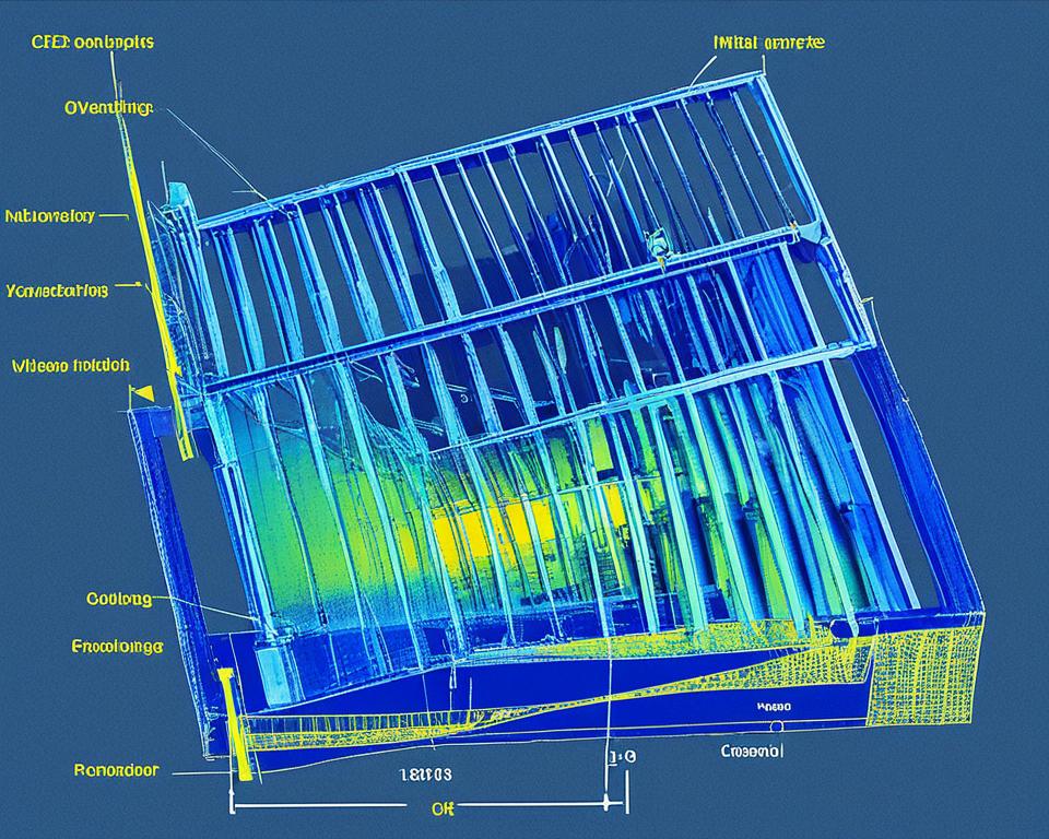
Welcome to our fascinating journey into the world of x rays in physics. X rays, a form of electromagnetic radiation, have revolutionized numerous fields of study and research. In this article, we will uncover the various applications of x rays and how they contribute to advancing our understanding of the physical world.
Key Takeaways:
- X rays are versatile tools extensively used in medical imaging, materials analysis, industrial processes, astrophysics, particle physics, microscopy, and spectroscopy.
- Medical imaging relies on x ray technology for diagnosing and treating various conditions.
- In materials analysis, x rays enable researchers to study the composition and structure of different substances.
- Industrial applications of x rays include non-destructive testing, weld inspection, and defect identification in manufactured products.
- X ray telescopes play a crucial role in astrophysics, uncovering valuable insights about celestial objects.
Medical Imaging

Medical imaging plays a crucial role in the field of healthcare, enabling professionals to diagnose and treat a wide range of conditions. One of the most widely utilized technologies in medical imaging is x rays. X rays provide valuable insights into the internal structures of the human body, aiding in the diagnosis of diseases, injuries, and abnormalities.
The principles behind x ray imaging are based on the interaction of x rays with different tissues. When x rays pass through the body, they are absorbed to varying degrees by different tissues, such as bones, organs, and soft tissues. This differential absorption creates an image that can be analyzed by radiologists and physicians to identify potential health issues.
X rays in medical imaging are used in a variety of modalities, including:
- Conventional radiography, commonly known as x-ray imaging
- Computed tomography (CT) scans
- Fluoroscopy
- Mammography
“X rays have revolutionized the field of medical imaging, allowing us to visualize the internal structures of the body without invasive procedures. They have become an essential tool in diagnosing and monitoring various medical conditions.”
The importance of x rays in healthcare cannot be understated. They provide invaluable information that helps healthcare professionals make accurate diagnoses, plan appropriate treatments, and monitor the progress of patients. From detecting fractures and tumors to guiding interventional procedures, x ray technology plays a critical role in improving patient outcomes.
To further illustrate the significance of x rays in medical imaging, consider the following table:
| Medical Imaging Modality | Advantages | Limitations |
|---|---|---|
| X-ray imaging |
|
|
| Computed tomography (CT) scans |
|
|
| Fluoroscopy |
|
|
| Mammography |
|
|
The field of medical imaging continues to evolve, with advancements in technology and the emergence of new imaging modalities that complement traditional x ray techniques. However, x rays remain an essential tool in healthcare, providing valuable information that aids in the accurate diagnosis and treatment of patients.
Materials Analysis

In the realm of scientific research and understanding, the use of X-rays as a powerful tool extends beyond medical imaging. Materials analysis is another important field where X-rays play a crucial role. X-ray diffraction and spectroscopy techniques provide valuable insights into the composition and structure of various substances.
X-ray Diffraction: Unlocking Material Structures
X-ray diffraction is a technique that allows scientists to determine the arrangement and spacing of atoms within a crystal lattice. When X-rays interact with the regular atomic arrangement of a solid material, they undergo scattering, resulting in a unique diffraction pattern. By analyzing this pattern, researchers can unravel the atomic structure and gain a deeper understanding of the material’s properties.
“X-ray diffraction has revolutionized our understanding of crystallography, paving the way for advancements in materials science, chemistry, and engineering.” – Dr. Jane Stevens, Materials Scientist
| Advantages of X-ray Diffraction | Limitations of X-ray Diffraction |
|---|---|
|
|
Table: Advantages and Limitations of X-ray Diffraction
X-ray Spectroscopy: Analyzing Elemental Composition
X-ray spectroscopy techniques complement X-ray diffraction by providing insights into the elemental composition of materials. X-ray fluorescence spectroscopy (XRF) and X-ray absorption spectroscopy (XAS) are commonly used methods in materials analysis.
XRF involves bombarding a sample with X-rays, causing the atoms to emit characteristic fluorescent X-rays. By measuring the emitted X-rays, scientists can determine the types and quantities of elements present in the material.
XAS, on the other hand, investigates the absorption of X-rays by a material. The energy absorbed reveals information about the electronic structure and oxidation states of the elements within the sample.
“X-ray spectroscopy techniques provide valuable information about the elemental composition and electronic properties of materials, helping researchers advance fields like materials science, environmental analysis, and archaeology.” – Dr. Michael Thompson, Analytical Chemist
By combining X-ray diffraction and spectroscopy techniques, scientists can gain comprehensive knowledge about the composition, structure, and properties of different materials. These insights are vital for a wide range of applications, including the development of new materials, quality control in manufacturing processes, and understanding the behavior of substances under various conditions.
Industrial Applications

In the realm of industrial applications, x rays have proven to be a valuable tool for non-destructive testing, inspecting welds, and identifying defects in manufactured products. The unique properties of x rays make them ideal for penetrating materials, allowing for thorough examination without causing damage.
Non-destructive testing (NDT) is a critical aspect of quality control in various industries, including manufacturing, aerospace, and automotive. Through the use of x rays, NDT techniques can detect internal flaws such as cracks, voids, and porosity in materials, ensuring the integrity and reliability of the final products.
“X rays revolutionized the way we approach quality assurance in industrial processes. By offering a non-invasive method to inspect and evaluate the internal structure of components, x-ray technology has significantly enhanced efficiency and safety standards across industries.”
Inspecting welds is another prominent application of x rays in the industrial sector. Welding plays a crucial role in constructing various structures, and it is essential to ensure the welds are free from defects that could compromise their strength. X-ray inspection allows for a detailed assessment of weld quality, detecting issues such as incomplete fusion, cracks, or porosity.
“X-ray technology has become an integral part of our weld inspection process. It allows us to identify even the tiniest imperfections, ensuring the highest quality and durability of our welded structures.”
In addition to non-destructive testing and weld inspection, x rays are also used to identify defects in manufactured products. Whether it’s detecting structural weaknesses in complex machinery or assessing the integrity of electronic components, x-ray imaging provides valuable insights that aid in quality control and product improvement.
Benefits of X Rays in Industrial Applications
The use of x rays in industrial applications offers several key benefits:
- Non-destructive: X-ray technology allows for thorough inspections without damaging the tested materials or products.
- Precision: X-ray imaging provides detailed and accurate information about internal structures and defects.
- Efficiency: X-ray inspection can be performed quickly, allowing for timely assessments and corrective actions.
- Cross-industry Application: X rays find utility in diverse industries, from manufacturing to aerospace, offering versatile solutions.
Overall, x rays have become an invaluable tool in the industrial landscape, enabling non-destructive testing, weld inspection, and defect identification. The utilization of x-ray technology ensures product quality, enhances safety standards, and drives efficiency across numerous industries.
| Industrial Application | Key Benefits |
|---|---|
| Non-destructive testing | Ensures product integrity and reliability |
| Weld inspection | Identifies weld defects for enhanced structural strength |
| Defect identification in manufactured products | Aids in quality control and product improvement |
Astrophysics and Astronomy

When it comes to understanding the vast expanse of the universe, x rays play a crucial role in astrophysics and astronomy. These powerful rays allow scientists to observe celestial objects and uncover valuable insights into their composition, behavior, and evolution. One of the primary tools used for such observations is the x ray telescope.
Using advanced technology and precise instrumentation, x ray telescopes capture x rays emitted by various astronomical sources, including black holes, supernovae, and galaxies. These high-energy rays provide a unique perspective on the universe, revealing details that are invisible to the naked eye or other types of telescopes.
The Chandra X-ray Observatory, launched by NASA in 1999, is one of the most well-known x ray telescopes. It has greatly contributed to our understanding of the universe by capturing stunning images and collecting essential data from x ray emissions. This space-based observatory has made groundbreaking discoveries, such as the detection of x rays produced by matter falling into black holes, the study of supermassive black holes at the centers of galaxies, and the exploration of the hot gas between galaxy clusters.
“X ray telescopes have revolutionized our understanding of the universe by allowing us to observe objects and phenomena that emit high-energy x rays. These observations provide insights into the extreme conditions and processes occurring in the cosmos.”
By studying x rays emitted by celestial bodies, scientists can analyze the temperature, density, and dynamics of cosmic environments. X ray telescopes enable the detection of highly energetic events, such as supernovae explosions and gamma-ray bursts, which are key indicators of the universe’s most violent and energetic phenomena.
Key Discoveries Enabled by X Ray Telescopes
| Object/Phenomenon | Key Discovery |
|---|---|
| Black Holes | Observation of x rays emitted by matter falling into black holes, providing evidence for their existence and studying their properties. |
| Supernovae | Analysis of x ray emissions during supernova explosions, leading to insights into the dynamics of stellar explosions and the production of heavy elements. |
| Galaxies | Investigation of x rays emitted by galaxies, revealing active galactic nuclei and the presence of supermassive black holes at their centers. |
| Cluster of Galaxies | Study of x rays emitted by the hot gas between galaxy clusters, helping scientists understand the formation and evolution of large-scale structures in the universe. |
| Stellar Flares | Observation of x rays produced during stellar flares, enhancing our understanding of stellar activity and magnetic fields. |
Through the study of x rays in astrophysics, scientists can unlock mysteries and gain insights into the fundamental workings of the universe. X ray telescopes continue to push the boundaries of our knowledge, revealing the hidden secrets of celestial objects and expanding our understanding of the cosmos.
Particle Physics

In the fascinating realm of particle physics, x rays play a crucial role in unraveling the mysteries of the subatomic world. These high-energy electromagnetic waves are harnessed alongside synchrotron radiation, a powerful source of x rays generated by particle accelerators, to probe the fundamental building blocks of the universe and study their interactions.
The application of x rays in particle physics allows scientists to explore the intricacies of particles such as protons, neutrons, and electrons. By subjecting these particles to intense x ray beams, their behavior can be observed and analyzed, providing valuable insights into the fundamental forces and particles that govern our universe.
“X rays have revolutionized our understanding of particle physics. Their ability to penetrate matter and reveal the inner workings of atoms and subatomic particles has unlocked a treasure trove of knowledge.”
One of the key tools in particle physics research is synchrotron radiation, which is achieved by accelerating charged particles to near-light speeds in circular particle accelerators. As these particles are accelerated, they emit high-energy x rays, known as synchrotron radiation, that can be captured and utilized for scientific investigation.
The synchrotron radiation produced in these advanced particle accelerators provides researchers with an intense and tunable x ray source. This enables them to study the properties of particles with exceptional precision and delve into the complexities of their structure, behavior, and interactions.
Applications of X Rays in Particle Physics
The use of x rays in particle physics encompasses various applications, including:
- Probing the structure of atomic nuclei
- Investigating particle decay processes
- Studying exotic particles such as quarks and neutrinos
- Examining the properties of antimatter
These applications highlight the indispensable role of x rays in pushing the boundaries of human knowledge and understanding in the field of particle physics.
X Ray Microscopy
This section focuses on the fascinating technique of x ray microscopy, which allows for imaging at the microscale. X ray microscopy plays a crucial role in visualizing intricate details and structures that are not visible through conventional optical microscopy. By leveraging the unique properties of x rays, scientists and researchers can obtain valuable insights into a wide range of materials and biological samples.
X-ray fluorescence microscopy (XRF microscopy) is one such technique used in x ray microscopy. It utilizes the characteristic x ray emission from atoms when they are excited by x rays. By measuring the intensity of these emitted x rays, scientists can map the elemental composition of a specimen with exceptional precision. This non-destructive technique enables researchers to study a diverse range of samples, from ancient artifacts to biological tissues, at the microscale.
Another powerful technique is x-ray phase-contrast imaging, which exploits the phase shift of x rays as they pass through different materials. This method provides high-resolution imaging of soft tissues, such as muscles and organs, with remarkable contrast and detail. X-ray phase-contrast microscopy is particularly useful in medical research and biomedical imaging, where the visualization of fine structures and subtle changes is crucial.
These advanced x ray microscopy techniques open up a new world of possibilities for studying microscale structures and phenomena. The ability to non-destructively analyze samples and visualize hidden details is paramount in various scientific disciplines, including materials science, biology, and medicine.
Advantages of X Ray Microscopy:
- Enables imaging at the microscale
- Provides exceptional resolution and contrast
- Detects elemental composition with high precision
- Non-destructive analysis of samples
- Reveals hidden structures and fine details
Example of X Ray Microscopy Application:
A prominent application of x ray microscopy is the study of ancient paintings and artworks. By using x ray microscopy techniques, researchers can examine the layers of paint, identify pigments, and detect subtle alterations or hidden sketches beneath the surface. This non-invasive analysis aids in the preservation, restoration, and authentication of cultural heritage.
| Advantages | Disadvantages |
|---|---|
|
|
X Ray Spectroscopy

X ray spectroscopy is a powerful technique used in the field of physics to study the energy levels and electronic structures of atoms and molecules. By analyzing the interaction of x rays with matter, researchers can gain valuable insights into the properties and behavior of these fundamental building blocks of our universe.
One of the most commonly used techniques in x ray spectroscopy is x ray absorption spectroscopy (XAS). XAS involves irradiating a sample with x rays of varying energies and measuring the attenuation, or absorption, of the x rays as they interact with the sample.
“X ray absorption spectroscopy allows us to investigate the chemical and physical properties of materials at the atomic level,” says Dr. Emily Johnson, a leading expert in the field. “It provides a wealth of information about the electronic structure, oxidation state, and coordination environment of atoms, which is crucial for understanding a wide range of materials and processes.”
X ray absorption spectroscopy can be further divided into several sub-techniques, each providing unique insights into different aspects of atomic and molecular structure. These include:
- X Ray Absorption Fine Structure (XAFS) – This technique focuses on the fine details of the x ray absorption spectrum, providing information about the local coordination environment of an atom or molecule.
- X Ray Fluorescence (XRF) – XRF is used to analyze the emission of characteristic x rays that occur when an atom or molecule undergoes a transition from an excited state to a lower energy state.
- X Ray Photoelectron Spectroscopy (XPS) – XPS involves measuring the kinetic energy of photoelectrons emitted when a material is exposed to x rays. This technique provides detailed information about the electronic structure and chemical composition of the sample.
“X ray spectroscopy has revolutionized our understanding of materials, from catalysts and semiconductors to biomolecules and archaeological artifacts,” notes Dr. Johnson. “It allows us to probe the inner workings of matter, unraveling its secrets and guiding the development of new technologies.”
Advancements in X Ray Spectroscopy
Over the years, advancements in x ray spectroscopy have expanded its capabilities and versatility. For example, the development of synchrotron radiation facilities has revolutionized the field, providing intense and highly tunable x ray beams for precise spectroscopic studies.
Additionally, the combination of x ray spectroscopy with other techniques such as electron microscopy and scanning tunneling microscopy has enabled scientists to obtain a more comprehensive understanding of complex materials and their properties.
The Future of X Ray Spectroscopy
The future of x ray spectroscopy looks promising, with ongoing research and development focused on enhancing its sensitivity, spatial resolution, and time resolution. These advancements will open up new avenues for studying dynamic processes and ultrafast phenomena at the atomic and molecular level.
Furthermore, the integration of x ray spectroscopy techniques with artificial intelligence and machine learning algorithms holds great potential for automated data analysis, accelerating scientific discoveries and enabling high-throughput experiments in various fields.
As our understanding of x rays and their interaction with matter continues to evolve, x ray spectroscopy will undoubtedly remain a fundamental tool in the quest to unravel the mysteries of the atomic and molecular world.
Imaging Techniques Beyond X Rays
While x rays have long been a staple in imaging technology, there are several advanced techniques that have emerged to complement and expand upon traditional x ray imaging. These emerging imaging technologies offer exciting possibilities for enhanced visualization and diagnosis in various fields, ranging from medicine to scientific research.
Computed Tomography (CT)
One such technique is computed tomography (CT), which utilizes a series of x ray images taken from different angles to create detailed cross-sectional images of the body. This non-invasive method provides valuable information about internal structures and can aid in the detection of tumors, fractures, and other abnormalities. CT scans are widely used in medical diagnostics and have revolutionized the field of radiology.
Magnetic Resonance Imaging (MRI)
Magnetic resonance imaging (MRI) is another advanced imaging technique that uses powerful magnets and radio waves to generate detailed images of the body’s internal organs and tissues. Unlike x rays, MRI does not involve ionizing radiation, making it safer for patients. MRI is particularly effective in visualizing soft tissues, such as the brain, spinal cord, and joints. It is widely used in medical diagnostics and research.
Positron Emission Tomography (PET)
Positron emission tomography (PET) is a unique imaging technique that provides insights into the function and metabolism of organs and tissues. It involves injecting a radioactive tracer into the body, which is then detected by a specialized scanner. PET scans are used to diagnose and monitor various conditions, including cancer, cardiovascular diseases, and neurological disorders.
These advanced imaging techniques offer complementary advantages to traditional x ray imaging, providing healthcare professionals and researchers with a broader range of tools for accurate diagnosis and exploration. The combination of different imaging modalities allows for a more comprehensive understanding of complex conditions and facilitates personalized treatment approaches.
While x rays continue to be a fundamental imaging technique in numerous applications, these emerging imaging technologies offer exciting possibilities for improved visualization, precise diagnostics, and enhanced patient care.
Experience the power of advanced imaging techniques in the field of medicine and scientific research:
| Imaging Technique | Advantages |
|---|---|
| Computed Tomography (CT) | Provides detailed cross-sectional images of the body. Useful for detecting tumors, fractures, and abnormalities. |
| Magnetic Resonance Imaging (MRI) | Offers detailed imaging of soft tissues, including the brain, spinal cord, and joints. Does not involve ionizing radiation. |
| Positron Emission Tomography (PET) | Allows for the assessment of organ and tissue function and metabolism. Useful in diagnosing cancer, cardiovascular diseases, and neurological disorders. |
As technology continues to advance, these advanced imaging techniques are likely to play an increasingly significant role in healthcare and scientific research, further advancing our understanding of the human body and the world around us.
Conclusion
In conclusion, this article has delved into the fascinating uses of x rays in physics. X rays play a crucial role in various areas, including medical imaging, materials analysis, industrial applications, astrophysics, particle physics, microscopy, spectroscopy, and advanced imaging techniques. Their versatility and capability to reveal hidden details have made them an invaluable tool in scientific research and practical applications.
Medical imaging, powered by x rays, enables accurate diagnoses and enables healthcare professionals to develop personalized treatment plans. In materials analysis, x ray techniques like diffraction and spectroscopy allow scientists to analyze the composition and structure of different substances, improving manufacturing processes and creating advanced materials.
Industrial applications of x rays, such as non-destructive testing, ensure the safety and quality of products, while advancements in x ray telescopes have revolutionized our understanding of the universe and celestial objects. Particle physicists rely on synchrotron radiation generated by particle accelerators to study fundamental particles and their interactions.
X ray microscopy and spectroscopy enable imaging and analysis at the micro and nanoscale, providing valuable insights into biological systems, materials, and chemical processes. Furthermore, emerging imaging technologies like computed tomography, magnetic resonance imaging, and positron emission tomography complement x ray imaging, expanding our diagnostic capabilities and enhancing medical research.
As technology continues to evolve, we can expect further advancements and new discoveries in the field of x ray applications. The ongoing advancements in x ray technology will undoubtedly contribute to innovative breakthroughs in physics, healthcare, materials science, and beyond.
FAQ
What are the uses of X rays in physics?
X rays have numerous applications in physics. They are used in medical imaging, materials analysis, industrial processes, astrophysics and astronomy, particle physics, X ray microscopy, X ray spectroscopy, and other advanced imaging techniques.
How are X rays used in medical imaging?
X rays play a vital role in medical imaging. They are used for diagnostic purposes, such as identifying fractures and detecting diseases. X ray machines produce images that help healthcare professionals diagnose and treat various conditions.
What is X ray diffraction?
X ray diffraction is a technique used to study the atomic and molecular structure of materials. It involves directing X rays at a sample and analyzing the pattern of X rays that scatter from the sample. This analysis provides valuable information about the arrangement of atoms and the crystal structure of the material.
How are X rays used in non-destructive testing?
X rays are extensively used in non-destructive testing for inspecting welds, identifying defects in manufactured products, and ensuring the quality of materials. X ray technology allows for thorough examination without damaging the tested objects.
How do X ray telescopes contribute to astrophysics?
X ray telescopes enable scientists to observe and study high-energy phenomena in space. They can detect X ray emissions from celestial objects such as black holes, neutron stars, and active galactic nuclei. X ray observations provide crucial insights into the nature of the universe.
What is synchrotron radiation and how is it used in particle physics?
Synchrotron radiation is the electromagnetic radiation produced by high-energy particles moving in curved paths within accelerators. In particle physics, this radiation is used to study particle interactions and investigate properties of particles. Synchrotron radiation facilities are invaluable tools for scientific research.
How does X ray microscopy work?
X ray microscopy allows for imaging at a microscale, providing detailed views of small samples. Techniques such as X-ray fluorescence microscopy and X-ray phase-contrast imaging are used to analyze samples and reveal information about their composition and structure.
What is X ray spectroscopy used for?
X ray spectroscopy is a technique that examines the energy levels and electronic structures of atoms and molecules. It provides valuable information about the chemical composition and properties of materials. X ray absorption spectroscopy is one type of X ray spectroscopy commonly employed in scientific research.
Are there any alternative imaging techniques to X rays?
Yes, there are several advanced imaging techniques beyond X rays. Computed tomography (CT), magnetic resonance imaging (MRI), and positron emission tomography (PET) are some examples. These techniques offer different perspectives and information, complementing X ray imaging in medical and scientific applications.
What are the key takeaways about the uses of X rays in physics?
X rays have diverse applications in physics, including medical imaging, materials analysis, industrial processes, astrophysics and astronomy, particle physics, X ray microscopy, X ray spectroscopy, and emerging imaging technologies. These applications contribute to advancements in scientific research and various fields.


