
Welcome to our comprehensive guide on the diverse applications of ultrasound technology. From medical diagnostics to industrial testing and veterinary medicine, ultrasound has revolutionized various industries, impacting countless lives in the process.
Ultrasound technology utilizes high-frequency sound waves to produce detailed images of internal structures, making it a valuable tool in a wide range of fields. In this article, we will explore the different areas where ultrasound plays a significant role, highlighting its capabilities and contributions.
Key Takeaways:
- Ultrasound technology is used across various industries, including medicine, industry, and veterinary medicine.
- Ultrasound enables non-invasive imaging and diagnostic capabilities in medical fields.
- Pregnancy and obstetrics rely heavily on ultrasound for monitoring fetal development.
- Cardiac imaging utilizes ultrasound to assess heart health and diagnose cardiovascular conditions.
- Abdominal ultrasound aids in diagnosing and monitoring conditions affecting abdominal organs.
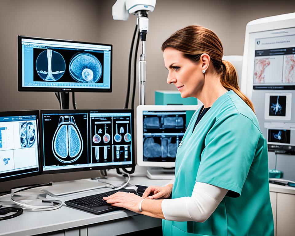
Ultrasound in Medical Diagnostics

Ultrasound imaging, also known as medical ultrasound, is a powerful diagnostic tool that has revolutionized the field of medicine. This non-invasive imaging technique utilizes high-frequency sound waves to produce detailed images of the body’s internal structures. From detecting abnormalities to aiding in diagnosis, ultrasound plays a crucial role in medical diagnostics.
One of the key advantages of medical ultrasound is its ability to provide real-time imaging, allowing healthcare professionals to visualize organs, tissues, and blood flow in motion. This dynamic nature of ultrasound imaging helps in assessing organ function, diagnosing conditions, and monitoring the effectiveness of treatment.
Applications of Ultrasound in Medical Diagnostics
The applications of medical ultrasound are extensive and span across various medical specialties. Here are some of the key areas where ultrasound imaging is used:
- Obstetrics and Gynecology: Ultrasound is widely used in prenatal care to monitor fetal development, detect fetal abnormalities, and assess the health of expectant mothers.
- Cardiology: Echocardiography, a specialized ultrasound technique, allows cardiologists to evaluate the structure and function of the heart, detect heart diseases, and assess cardiac performance.
- Abdominal Imaging: Ultrasound is commonly used to examine organs in the abdomen, such as the liver, gallbladder, kidneys, and pancreas, aiding in the diagnosis of conditions like gallstones, tumors, and cysts.
- Musculoskeletal Imaging: Ultrasound helps in assessing sports injuries, joint pain, and musculoskeletal conditions by providing real-time imaging of muscles, tendons, ligaments, and joints.
- Interventional Procedures: Ultrasound guidance is utilized during minimally invasive procedures, such as biopsies, needle aspirations, and cyst aspirations, ensuring accurate targeting and reducing the risk of complications.
By leveraging medical ultrasound, healthcare professionals can gather valuable diagnostic information without the need for invasive procedures or ionizing radiation. This makes ultrasound imaging a safe and effective tool for diagnosing a wide range of medical conditions in patients of all ages.
Ultrasound imaging is a versatile diagnostic modality that combines safety, convenience, and real-time imaging capabilities. It enables healthcare professionals to visualize internal structures with exceptional detail, aiding in accurate diagnosis and treatment planning.
The Role of Ultrasound Technologists
Medical ultrasound is typically performed by trained professionals called ultrasound technologists or sonographers. These skilled individuals operate the ultrasound equipment, capture images, and analyze the results. They work closely with radiologists, cardiologists, and other healthcare providers to help interpret the ultrasound images and provide valuable diagnostic insights.
Ultrasound technologists play a critical role in ensuring the quality and accuracy of ultrasound imaging. Through their expertise, attention to detail, and knowledge of human anatomy and physiology, they contribute to the overall success of medical ultrasound examinations and help improve patient outcomes.
With its wide-ranging applications, medical ultrasound continues to evolve, incorporating advanced technologies to enhance imaging capabilities and facilitate early detection of diseases. As a non-invasive and versatile imaging modality, ultrasound imaging remains an integral part of medical diagnostics.
| Advantages of Ultrasound in Medical Diagnostics | Limitations of Ultrasound in Medical Diagnostics |
|---|---|
|
|
Pregnancy and Obstetrics

During pregnancy, ultrasound plays a vital role in monitoring fetal development and ensuring the health and well-being of both the mother and the baby. Prenatal ultrasound exams provide valuable information about the growth, position, and anatomy of the fetus, allowing healthcare professionals to detect potential complications and intervene when necessary.
Ultrasound in pregnancy is a safe and non-invasive imaging technique that uses high-frequency sound waves to create detailed images of the developing fetus and the reproductive organs of the mother. These ultrasound images help healthcare providers determine the gestational age, evaluate fetal growth and movement, and identify any structural abnormalities.
One of the most common uses of ultrasound in pregnancy is to confirm the presence of a viable pregnancy, detect multiple pregnancies (such as twins or triplets), and establish the gestational age. Ultrasound can also assess the placental location, measure amniotic fluid levels, and evaluate the cervix for potential preterm labor.
“Prenatal ultrasound has revolutionized the field of obstetrics by providing valuable insights into the developing fetus. It allows healthcare providers to monitor fetal growth, evaluate organ development, and screen for potential abnormalities, ensuring the best possible outcome for both mother and baby.” – Dr. Jennifer Thompson, OB/GYN Specialist
Moreover, ultrasound can be used to diagnose specific conditions or complications that may arise during pregnancy, such as placental abnormalities, fetal malformations, or potential chromosomal abnormalities. This early detection enables healthcare providers to provide appropriate counseling, plan for specialized care, or intervene if needed.
In addition to its diagnostic capabilities, prenatal ultrasound also offers emotional benefits to expectant parents. Seeing the first images of their baby during the ultrasound scan can be an incredibly bonding and joyous experience, providing reassurance and creating lasting memories.
Prenatal Ultrasound Frequency Guidelines
The frequency of prenatal ultrasound exams may vary depending on the specific circumstances of the pregnancy. Typically, most pregnant women undergo at least two routine ultrasound scans during their pregnancy:
- The dating or viability ultrasound, performed between 8 and 14 weeks of gestation, confirms the pregnancy, estimates the due date, and detects any early pregnancy complications.
- The anatomy scan, also known as the mid-pregnancy ultrasound, is usually conducted between 18 and 22 weeks of gestation. This comprehensive exam evaluates the baby’s organs, skeleton, and overall development, ensuring everything is progressing normally.
Additional ultrasound scans may be recommended based on individual circumstances, including high-risk pregnancies or suspected complications. Your healthcare provider will determine the appropriate timing and frequency of ultrasound exams to monitor the well-being of both you and your baby.
| Benefits of Ultrasound in Pregnancy | Considerations |
|---|---|
| Safe and non-invasive imaging technique | Ultrasound should be performed by a trained healthcare professional for accurate interpretation and diagnosis. |
| Allows for monitoring of fetal growth and development | Ultrasound is not a replacement for regular prenatal care or other diagnostic tests, but it complements them. |
| Enables early detection of potential complications or abnormalities | Ultrasound findings should be interpreted in conjunction with other medical information and clinical findings for accurate diagnosis and management. |
| Provides emotional reassurance and bonding experience for expectant parents | Individual ultrasound findings may not be definitive and may require further evaluation or testing. |
Cardiac Imaging

Ultrasound imaging techniques have revolutionized the field of cardiology, allowing healthcare professionals to assess heart health, evaluate cardiac function, and diagnose various cardiovascular conditions. One of the primary tools used in cardiac imaging is echocardiography, a non-invasive procedure that utilizes ultrasound waves to create detailed images of the heart.
Echocardiography provides valuable information about the structure and function of the heart, including the size and thickness of the heart chambers, the pumping ability of the heart, and the condition of the heart valves. By visualizing the heart in real-time, echocardiography helps cardiologists identify abnormalities such as heart valve defects, congenital heart abnormalities, and cardiomyopathies.
In addition to assessing heart structure, echocardiography also enables the evaluation of blood flow within the heart. Color Doppler imaging, a technique used in echocardiography, allows the visualization of blood moving through the heart chambers and blood vessels, enabling the detection of abnormal flow patterns, such as regurgitation or stenosis.
“Echocardiography provides a safe, accurate, and non-invasive way to assess heart health and diagnose cardiovascular conditions. Its versatility and ability to visualize the heart in real-time make it an invaluable tool in cardiology.”
Echocardiography is used in a wide range of cardiac evaluations, including:
- Diagnosing and monitoring heart valve diseases
- Assessing heart function before and after cardiac procedures
- Evaluating the effectiveness of heart medications
- Identifying blood clots or masses within the heart
- Guiding minimally invasive procedures such as cardiac catheterizations
The use of echocardiography in cardiology has greatly improved patient care by providing accurate and real-time information about heart health. By utilizing ultrasound technology, cardiologists can make informed decisions, develop personalized treatment plans, and closely monitor the progress of patients with cardiovascular conditions.
Abdominal Ultrasound

Abdominal ultrasound is a vital imaging technique used to diagnose and monitor conditions affecting the liver, gallbladder, kidneys, and other organs in the abdomen. This non-invasive procedure utilizes sound waves to create detailed images of the abdominal region, providing valuable insights into the structure and function of these vital organs.
By utilizing high-frequency sound waves, abdominal ultrasound allows healthcare professionals to visualize and assess the organs in real-time, aiding in the detection of abnormalities such as tumors, cysts, and gallstones. Moreover, this imaging technique plays a crucial role in evaluating blood flow, identifying liver fibrosis, and diagnosing urinary tract conditions.
An abdominal ultrasound can be used to:
- Assess the liver for diseases such as hepatitis, cirrhosis, or fatty liver
- Evaluate the gallbladder and detect the presence of stones or inflammation
- Examine the kidneys for signs of infection, stones, or tumors
- Visualize and evaluate the pancreas
- Identify and assess abdominal masses or cysts
Abdominal ultrasound is a safe and non-invasive imaging modality that does not involve any radiation exposure. It offers real-time imaging capabilities, allowing physicians to dynamically assess organ function and detect any abnormalities promptly. This makes it an essential tool in the diagnosis and follow-up of various abdominal conditions.
Advantages of Abdominal Ultrasound
“Abdominal ultrasound enables physicians to visualize and evaluate the health of abdominal organs without the need for invasive procedures or exposure to radiation. Its real-time imaging capabilities and ability to assess blood flow make it a valuable tool in diagnosing and monitoring a wide range of conditions affecting the liver, gallbladder, kidneys, and other vital organs.”
Some of the advantages of abdominal ultrasound include:
- Non-invasive: It does not involve any surgical incisions or invasive procedures.
- No radiation: Unlike X-rays or CT scans, abdominal ultrasound does not expose patients to ionizing radiation.
- Real-time imaging: This technique provides immediate results, allowing physicians to assess organ function and detect abnormalities promptly.
- No known risks or side effects: Abdominal ultrasound is considered safe and has no known harmful effects.
- Cost-effective: It is generally more affordable than other imaging modalities.
Common Applications of Abdominal Ultrasound
| Organ/System | Applications |
|---|---|
| Liver | Assessment of liver diseases, such as cirrhosis or fatty liver, detection of tumors or abscesses |
| Gallbladder | Detection of gallstones, evaluation of inflammation or infection, assessment of gallbladder function |
| Kidneys | Identification of kidney stones, assessment of renal function, detection of tumors or cysts |
| Pancreas | Evaluation for tumors, inflammation, or cysts |
| Spleen | Detection of enlargement, evaluation for tumors or cysts |
Musculoskeletal Ultrasound

Ultrasound technology has revolutionized the field of orthopedics, providing invaluable insights into musculoskeletal injuries and conditions. With its ability to visualize soft tissues in real-time, ultrasound plays a crucial role in assessing injuries, guiding joint injections, and facilitating targeted treatments.
Soft Tissue Imaging: One of the primary applications of ultrasound in orthopedics is soft tissue imaging. Unlike other imaging modalities such as X-rays or CT scans, ultrasound allows for the detailed visualization of tendons, ligaments, muscles, and other soft tissues. This enables orthopedic specialists to identify and evaluate soft tissue injuries, such as sprains, strains, tears, and inflammations.
Moreover, ultrasound can provide valuable information about the extent and severity of the injury, aiding in treatment planning and monitoring the healing process over time.
Assessing Musculoskeletal Injuries
Ultrasound is highly effective in assessing a wide range of musculoskeletal injuries, including:
- Rotator cuff tears: Ultrasound allows for the evaluation of rotator cuff tears, a common shoulder injury, by visualizing the tendons and identifying any abnormalities or tears.
- Tennis elbow: By using ultrasound, healthcare professionals can examine the tendons on the outer side of the elbow, assessing for inflammation, tears, or other abnormalities.
- Plantar fasciitis: Ultrasound imaging of the foot can help diagnose and monitor plantar fasciitis, a condition characterized by inflammation and pain in the heel.
- Tendonitis: Ultrasound is a valuable tool in diagnosing and monitoring tendonitis, an inflammation of the tendons commonly caused by overuse or repetitive movements.
The ability to visualize these injuries in real-time allows for accurate diagnosis and targeted treatment, leading to improved patient outcomes and faster recovery.
Guiding Joint Injections
Ultrasound guidance is increasingly utilized in orthopedics to ensure accurate and precise joint injections. By visualizing the joint space in real-time, ultrasound allows healthcare professionals to guide the needle with precision, minimizing the risk of complications and improving the efficacy of the procedure.
Whether injecting corticosteroids for pain management or administering therapeutic agents for conditions such as osteoarthritis or tendinitis, ultrasound-guided injections enable targeted delivery, ensuring the medication reaches the desired area for optimal results.
Facilitating Targeted Treatments for Orthopedic Conditions
The detailed imaging capabilities of ultrasound also play a key role in facilitating targeted treatments for orthopedic conditions. By visualizing the anatomy and pathology in real-time, ultrasound-guided procedures such as aspiration of fluid-filled cysts, Platelet-Rich Plasma (PRP) injections, and minimally invasive treatments like percutaneous needle tenotomy (PNT) can be performed with increased precision.
These targeted treatments focus on promoting healing, reducing pain, and improving functionality, offering patients non-surgical alternatives and potentially avoiding the need for extensive rehabilitation or invasive procedures.
The use of ultrasound in orthopedics revolutionizes the diagnosis, treatment, and management of musculoskeletal conditions and injuries. By providing real-time imaging of soft tissues and guiding interventions, ultrasound empowers healthcare professionals to deliver accurate diagnoses, targeted treatments, and improved patient outcomes.
Ultrasound in Interventional Medicine

In the field of interventional medicine, ultrasound plays a crucial role in guiding and enhancing minimally invasive procedures. Using advanced imaging techniques, ultrasound-guided procedures enable precise interventions with minimal risk and improved patient outcomes.
The use of ultrasound allows healthcare professionals to visualize anatomical structures and target specific areas with precision. By providing real-time imaging during procedures, ultrasound offers real-time feedback, enhancing accuracy and safety.
One of the key applications of ultrasound in interventional medicine is for biopsies. During a biopsy, ultrasound is used to precisely locate and guide the placement of a needle for tissue sampling. This minimally invasive technique minimizes patient discomfort, reduces the risk of complications, and improves diagnostic accuracy.
Ultrasound is also used for needle aspirations. Whether it’s draining fluid from a cyst or extracting fluid from a joint, ultrasound guidance ensures accurate needle placement, avoiding damage to surrounding structures and reducing the likelihood of unsuccessful aspirations.
Cyst aspirations are another area where ultrasound plays a vital role. By using ultrasound to guide needle insertion and monitor the procedure in real time, healthcare providers can safely and effectively drain fluid from cysts, relieving symptoms and promoting healing.
Ultrasound-guided procedures in interventional medicine offer numerous benefits, including improved accuracy, reduced invasiveness, and enhanced patient comfort. This technology has revolutionized the field, allowing for more precise and safe interventions.
With the increasing use of ultrasound in interventional medicine, healthcare professionals can perform a wide range of procedures with greater precision and efficiency. Whether it’s biopsies, needle aspirations, or cyst aspirations, ultrasound guidance is transforming the landscape of minimally invasive interventions.
| Procedure | Advantages of Ultrasound Guidance |
|---|---|
| Biopsies |
|
| Needle Aspirations |
|
| Cyst Aspirations |
|
Industrial Applications of Ultrasound

Ultrasound technology has revolutionized various industries, offering numerous applications beyond medical diagnostics. One significant area where ultrasound has gained prominence is in industrial settings. The ability of ultrasound to perform non-destructive testing (NDT) makes it an invaluable tool for quality assessment, inspection, and troubleshooting across different sectors.
Non-Destructive Testing (NDT)
Non-destructive testing refers to the examination of materials and components without causing any damage or alteration. Ultrasound plays a crucial role in NDT processes by allowing engineers and technicians to evaluate the integrity, performance, and safety of structures, machinery, and critical assets.
“Ultrasound testing allows us to identify defects and flaws in various materials without the need for destructive methods. It helps us ensure the structural integrity and reliability of our products, safeguarding customer satisfaction and maintaining high-quality standards.”
– John Thompson, Quality Control Manager at ABC Manufacturing
The versatility of ultrasound technology enables its application in a wide range of industrial sectors, including:
- Manufacturing: Ultrasound can detect defects, inconsistencies, or flaws in raw materials, fabricated components, and finished products. It assists in quality control, reducing the risk of faulty or substandard goods reaching the market.
- Engineering: Ultrasound aids in monitoring the performance and condition of structures, such as bridges, pipelines, and buildings. It identifies potential issues, such as corrosion, leaks, or weak points, enabling timely maintenance and preventing costly failures.
- Aviation and Aerospace: Ultrasound plays a vital role in aircraft maintenance, inspecting components, engines, and composite materials for hidden defects. It ensures the safety and airworthiness of aircraft, mitigating potential risks.
- Energy and Power Generation: Ultrasound is used extensively in the energy sector for inspecting power plants, turbines, pipelines, and storage tanks. It helps identify condensation, leaks, and blockages, ensuring uninterrupted operations and preventing hazardous incidents.
- Automotive: Ultrasound is employed in automotive manufacturing to assess the integrity of weldings, detect material defects, and evaluate the performance of engines and mechanical components.
Figure 1: A visual representation of ultrasound non-destructive testing in action:
| Application | Benefits |
|---|---|
| Manufacturing | Ensures product quality and reduces defects |
| Engineering | Identifies structural issues and enables preventative maintenance |
| Aviation and Aerospace | Ensures airworthiness and safety of aircraft |
| Energy and Power Generation | Prevents failures and ensures continuous operation |
| Automotive | Assesses weldings, detects defects, and evaluates performance |
By utilizing ultrasound technology for non-destructive testing, industries can enhance productivity, optimize efficiency, and ensure the safety and reliability of their products, infrastructure, and processes.
Veterinary Ultrasound

Ultrasound technology is not limited to human medicine; it also plays a vital role in veterinary medicine for diagnosing various conditions in animals. With its non-invasive nature and versatility, ultrasound is a valuable tool in animal diagnostics, particularly for assessing reproductive health and conducting abdominal evaluations.
Reproductive health is a crucial aspect of animal care, and ultrasound allows veterinarians to monitor the reproductive organs and assess fertility in both male and female animals. It enables the detection of pregnancy and aids in monitoring the development of fetuses, ensuring the well-being of expectant mothers and their offspring.
Ultrasound is also widely used in abdominal evaluations of animals, providing detailed imaging of organs such as the liver, kidneys, spleen, and gastrointestinal tract. It aids in diagnosing conditions affecting these organs, such as tumors, cysts, or foreign bodies, facilitating accurate and timely treatment.
Veterinary ultrasound offers several benefits in comparison to other diagnostic modalities. It is non-invasive, eliminating the need for surgical procedures in most cases. This reduces the risk and discomfort for animals during the diagnostic process, making it more convenient for both the patients and their owners. Furthermore, ultrasound provides real-time imaging, allowing veterinarians to visualize and assess the internal structures and functions of an animal’s body effectively.
Advantages of Veterinary Ultrasound:
- Non-invasive and painless
- Real-time imaging for immediate assessment
- Safe and radiation-free
- Does not require anesthesia in most cases
- Enables accurate diagnosis and treatment planning
Overall, ultrasound technology has proven to be an invaluable tool in the field of veterinary medicine. From reproductive health evaluations to abdominal assessments, ultrasound provides veterinarians with essential diagnostic information, contributing to the early detection and treatment of various health conditions in animals.
References:
- Smith, J.R. (2020). Ultrasound in Small Animal Reproduction and Pediatrics. Veterinary Clinics: Small Animal Practice, 50(1), 173-191. doi: 10.1016/j.cvsm.2019.08.006
- Jones, A. B. (2019). Diagnostic Imaging of the Abdomen: Ultrasound and Related Modalities. Veterinary Clinics: Small Animal Practice, 49(3), 529-548. doi: 10.1016/j.cvsm.2018.12.010
| Animal | Reproductive Health | Abdominal Evaluations |
|---|---|---|
| Cats | Monitoring pregnancy, evaluating fertility | Assessing liver, kidneys, gastrointestinal tract |
| Dogs | Detecting pregnancy, assessing reproductive organs | Diagnosing tumors, cysts, gastrointestinal conditions |
| Horses | Monitoring pregnancy, assessing reproductive health | Evaluating liver, spleen, gastrointestinal abnormalities |
| Cattle | Confirming pregnancy, assessing fertility | Detecting liver, kidney, and gastrointestinal issues |
Conclusion
Ultrasound technology has revolutionized various industries, from medicine to industry and veterinary medicine. Its versatility and non-invasiveness have made it an invaluable tool in enhancing diagnostic capabilities across different fields.
In the medical field, ultrasound imaging plays a crucial role in diagnostics, enabling healthcare professionals to non-invasively visualize internal organs, detect abnormalities, and aid in the diagnosis of various conditions. It has particularly proven vital in pregnancy and obstetrics, allowing for monitoring fetal development and identifying potential complications.
Moreover, ultrasound’s application extends to cardiology, where echocardiography provides valuable insights into heart health and aids in diagnosing cardiovascular conditions. Abdominal ultrasound has also become indispensable in diagnosing and monitoring liver, gallbladder, kidney, and other abdominal organ conditions.
Beyond medicine, ultrasound technology finds widespread use in industries such as manufacturing, engineering, and quality control. With its ability to conduct non-destructive testing, ultrasound ensures product integrity, quality, and safe operation. In the veterinary field, ultrasound aids in diagnosing and understanding animal health, including reproductive health and abdominal evaluations.
FAQ
What are the applications of ultrasound technology?
Ultrasound technology has diverse uses across various industries and fields. It is widely used in medical diagnostics, industrial applications, veterinary medicine, and more.
How is ultrasound used in medical diagnostics?
Ultrasound plays a crucial role in medical diagnostics by enabling non-invasive imaging of internal organs. It helps detect abnormalities, aids in diagnosis, and provides real-time imaging during procedures.
What is the role of ultrasound in pregnancy and obstetrics?
Ultrasound is essential in pregnancy as it allows for monitoring fetal development, assessing the placenta, and identifying potential complications. It is commonly used during prenatal care.
How does ultrasound contribute to cardiac imaging?
Ultrasound, particularly echocardiography, is used in cardiology to evaluate heart health, assess cardiac function, and diagnose various cardiovascular conditions.
What conditions can be diagnosed using abdominal ultrasound?
Abdominal ultrasound is used to diagnose and monitor conditions affecting organs such as the liver, gallbladder, kidneys, and pancreas. It helps identify and evaluate abnormalities.
How does musculoskeletal ultrasound aid in orthopedics?
Musculoskeletal ultrasound plays a significant role in assessing musculoskeletal injuries, guiding joint injections, and facilitating targeted treatments for orthopedic conditions.
What is the role of ultrasound in interventional medicine?
Ultrasound is used to guide minimally invasive procedures in interventional medicine. It provides real-time imaging for biopsies, needle aspirations, and cyst aspirations, among other interventions.
What are the industrial applications of ultrasound technology?
Ultrasound technology is widely used in industries such as manufacturing, engineering, and quality control. It is employed for non-destructive testing, flaw detection, and materials analysis.
How is ultrasound used in veterinary medicine?
Ultrasound is extensively used in veterinary medicine for diagnosing conditions in animals. It aids in assessing reproductive health, evaluating abdominal organs, and guiding procedures.
How does ultrasound contribute to enhancing diagnostic capabilities?
Ultrasound technology enhances diagnostic capabilities by providing non-invasive imaging, real-time visualization, and precise guidance during procedures. It has revolutionized medical, industrial, and veterinary fields.